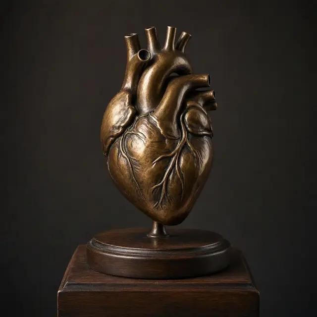
A Fist-Sized Pump
The heart is a muscular organ found in humans and other animals, which pumps blood through the blood vessels. Together they make up the circulatory system.
The job is delivery and collection at once: the pumped blood carries oxygen and nutrients to the tissues, while carrying metabolic waste such as carbon dioxide to the lungs.
In humans it is approximately the size of a closed fist, sitting between the lungs in the middle compartment of the chest, called the mediastinum. It is divided into four chambers — the upper left and right atria, and the lower left and right ventricles. The right atrium and ventricle are usually referred to together as the right heart, and their left counterparts as the left heart. It is enclosed in a protective sac, the pericardium, and its wall has three layers: epicardium, myocardium and endocardium.
It Generates Its Own Signal
The most remarkable thing about the heart is that nothing tells it to beat.
It pumps with a rhythm determined by a group of pacemaker cells in the sinoatrial node. These generate an electric current that causes the heart to contract, travelling through the atrioventricular node and along the heart's own conduction system.
The heart contains its own electrical generator and its own wiring. The brain can speed it up or slow it down, but the beat originates inside the muscle itself — which is why a heart can continue beating outside the body under the right conditions, and why the organ is described as having its own rhythm rather than receiving one. In adults, the resting rate is close to 72 beats per minute. Blood flows one way only, because heart valves prevent backflow.
The Route Blood Takes
The circuit runs in a figure of eight, and it passes through the heart twice.
Deoxygenated blood enters the right atrium from the superior and inferior venae cavae, and passes to the right ventricle. From there it is pumped into the pulmonary circulation to the lungs, where it takes on oxygen and gives off carbon dioxide.
Oxygenated blood then returns to the left atrium, passes through the left ventricle, and is pumped out through the aorta into the systemic circulation. It travels through arteries, then arterioles, then capillaries — where nutrients and other substances are exchanged between vessels and cells — before returning through venules and veins to start again. That is why the left side is more muscular: it pushes blood around the entire body, while the right side only has to reach the lungs.
The Leading Cause of Death
Cardiovascular diseases are the most common cause of death globally. An estimated 19.8 million people died from them in 2022 — about 32% of all deaths worldwide. Of those, 85% were due to heart attack and stroke.
The risk factors are largely known and largely shared: smoking, being overweight, little exercise, high cholesterol, high blood pressure, and poorly controlled diabetes, among others.
The difficult part is detection. Cardiovascular diseases do not frequently have symptoms, though they may cause chest pain or shortness of breath. Diagnosis is often made by taking a medical history, listening to heart sounds with a stethoscope, and using an ECG or an echocardiogram, which uses ultrasound. Specialists in the heart are called cardiologists, though many medical specialties may be involved in treatment.